Researchers from Trinity College Dublin in Ireland have discovered a human receptor that is critical in blood clotting and inflammation. In doing so, they created a “molecular map” that could influence more effective therapies for people who have pulmonary arterial hypertension (PAH), lung fibrosis, cardiovascular disease or certain cancers.

The paper, “Structural and Dynamic Insights Into Agonist Recognition and Function of the Thromboxane A2 Receptor,” was published in Nature Communications.
The identified receptor, thromboxane A2, appears on blood platelets and other cell types, regulating inflammatory responses, blood vessel contraction and blood clot formation. Using advanced cryo-election microscopy, the team captured high-res images of the receptor while it was actively transmitting signals across the membrane.
“Thromboxane A2 itself is a short-lived signaling molecule that disappears within seconds in the body, which has always made it difficult for us to study how it activates the receptor,” said Pawel Krawinski, PhD, in a university news release. Dr. Krawinski is a postdoctoral research fellow in Trinity College Dublin’s School and Medicine and School of Biochemistry and Immunology.
“To overcome this, we used a new technique which allowed us to visualize its structure in extraordinary detail, and that in turn helped us learn how it interacts with signaling proteins inside the cell,” he said.
According to the researchers, the structural images revealed surprising information. Thromboxane A2, unlike similar receptors, utilizes an unusual “activation switch” to trigger internal signaling. The team also discerned that signaling molecules appeared to enter the receptor from within the cell membrane rather than from outside the cell.

“These insights are fascinating to us, but they are far more than just structural details, as they could have important medical implications too,” Dr. Krawinski said. “The thromboxane receptor plays a role in multiple diseases, including cardiovascular and cardiopulmonary disorders, pulmonary arterial hypertension and fibrotic lung disease. It is also overactive in some cancers and inflammatory conditions.”
The authors noted the study also provides critical information on bleeding disorders caused by rare inherited mutations in the thromboxane receptor. By understanding how the mutations affect the receptor’s structure, researchers could develop improved diagnostic methods and treatments for patients with these diseases, they said.
“By revealing how activating molecules bind and trigger the receptor, the new molecular map provides a blueprint for developing drugs that can selectively block or fine-tune its activity,” Dr. Krawinski said. “Such drugs could help prevent harmful blood clotting, reduce excessive blood vessel constriction or limit inflammatory signaling linked to disease.”
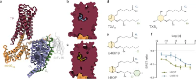
*a Cartoon representation of the active TP structures bound to the agonist I-BOP and the mini-Gq heterotrimer. The TP is coloured dark red, with mini-Gαq in gold, Gβ and Gγ in purple and green, respectively, and scFv16 in light grey. I-BOP is in olive green. Cutaway view of the U46619 (b, blue) and I-BOP (c, green) bound TP structures indicating that both ligands adopt an L-shaped conformation, while occupying a binding pocket that is buried deep within the receptor’s transmembrane core. The sealed cavity is proposed to protect the ligands from decomposition by hydration to the water-soluble TXB2. dChemical structures of the endogenous TP agonist TXA2 (left) and its derivative TXB2 (right). e Chemical structures of the synthetic agonists used in this study: U46619 and I-BOP. In (d) and (e), the α- and ω-chains are labelled, the bicyclic ring that differs between the synthetic agonists and the endogenous TXA2 is in yellow, and the iodobenzene group present in I-BOP is in green. f Gq heterotrimer dissociation as measured by a loss of BRET after TP stimulation with U46619 (blue) or I-BOP (green) in HEK293 cells transiently co-expressing the wild-type receptor and TRUPATH biosensors for the full-length Gq protein. Data are represented as mean ± SEM from 8 independent experiments for U46619 and 4 independent experiments for I-BOP, performed in triplicate, indicating EC50 values of 11.22 ± 5.29 nM and 1.79 ± 1.08 nM (mean ± SEM) for U46619 and I-BOP, respectively.