Researchers have developed an AI model that predicts pneumonitis risk in lung cancer patients before immunotherapy treatment begins.
- CIPHER is an AI-based model that detects imaging patterns in routine CT scans to predict a person’s risk of developing pneumonitis — a potentially life-threatening side effect.
- The model achieved an area under the curve (AUC) of 0.83, outperformed traditional approaches that rely on clinical risk factors and subjective imaging analysis.
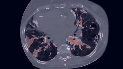
- The model identifies subtle abnormalities in lung tissue rather than looking for pneumonitis itself, revealing hidden information in routine imaging.
People who undergo treatment for lung cancer are at risk of developing pneumonitis, a life-threatening side effect associated with immunotherapy. Now, an artificial intelligence (AI)-based model can predict pneumonitis risk before treatment begins.
Researchers at The University of Texas MD Anderson Cancer Center developed the model, called Checkpoint-Inhibitor Pneumonitis Hazard Estimator (CIPHER). According to lead investigator Jia Wu, PhD, CIPHER analyzes routine chest CT scans to detect imaging patterns that indicate a patient’s risk of developing pneumonitis. Dr. Wu is an associate professor of imaging physics and thoracic/head and neck medical oncology and an affiliate member of UT MD Anderson’s Institute for Data Science in Oncology in Houston.

“Pneumonitis remains one of the most challenging complications of immunotherapy because it can be difficult to predict before symptoms appear,” said Dr. Wu in an MD Anderson news release. “Our model was able to identify signals associated with future risk using information that already exists in routine CT scans.”
 Jia Wu, PhD
The scientists built CIPHER using more than 590,000 CT image slices from 2,500 patients with lung cancer. First, they trained the model to recognize lung tissue patterns found in imaging. From there, the model analyzed the patterns to determine a person’s risk of developing pneumonitis after immunotherapy treatment.
Next, researchers tested the AI-based model with pretreatment CT scans from 347 patients who were treated for non-small cell lung cancer (NSCLC) at UT MD Anderson. They also validated the model using an independent external dataset.

In both experiments, CIPHER attained an area under the curve (AUC) of about 0.83. This indicates the model’s predictive power, which outperformed traditional approaches that rely on clinical risk factors and subjective imaging analysis, the researchers said.
“What makes this approach particularly interesting is that it was not designed to look for pneumonitis itself,” Dr. Wu said. “Instead, the model learned patterns within lung tissue and identified subtle abnormalities associated with future risk. That suggests routine imaging may contain much more information about treatment toxicity than we previously recognized.”
CIPHER demonstrated strong predictive performance throughout the experiments, the researchers noted, despite inconsistencies in patient populations, CT scanners and imaging protocols. The model also adjusted for factors like age, smoking history, tumor histology and prior radiation exposure.
Dr. Wu said larger studies with more diverse patient populations are needed before CIPHER can be clinically implemented. The researchers said they also plan to assess the model’s performance in other types of cancer that are commonly treated with immunotherapy.