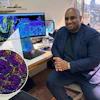
A novel computer model of a human lung can simulate how a burst of radiation interacts with the organ on a cell-by-cell level. Scientists from the University of Surrey and GSI Helmholtzzentrum für Schwerionenforschung, Darmstadt, developed the model using artificial intelligence (AI). The team’s research, published in Communications Medicine, found that the model can help improve radiotherapy treatment by making it safer and more accurate, thereby reducing the potential damage. It could also lead to more targeted treatments for other types of cancer.

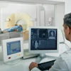
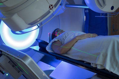
Currently, more than half of lung cancer patients receive radiotherapy, but too high a dose can injure their lungs. This can lead to the development of secondary conditions, such as pneumonitis and fibrosis.
"Doctors could one day use our model to choose the right length and strength of radiotherapy—tailored to their patient,” said Roman Bauer, MCSE, senior lecturer at the University of Surrey. “This is exciting enough—but others could use our technique to study other organs. This could unlock all kinds of medical knowledge and could be great news for doctors and future patients."
"For the first time, BioDynaMo makes interactive models of entire human organs achievable,” said Marco Durante, PhD, professor and head of the biophysics department at GSI. “This will allow us to model individual patients' lungs in a way that's just not possible with the very general statistical methods we currently use. What's more—it will allow us to study the way fibrosis and other conditions are actually caused, and how they develop over time."