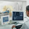
A new positron emission tomography (PET) imaging technique can accurately detect and monitor difficult-to-diagnose lung infections, particularly Mycobacteroides abscessus. Researchers said the novel diagnostic tool could significantly improve diagnosis and treatment for patients with chronic lung infections. The abstract, “Dynamic 11C-PABA PET/CT for Visualizing Pulmonary Mycobacteroides Abscessus Infections,” was presented at the 2025 Society of Nuclear Medicine and Molecular Imaging (SNMMI) Annual Meeting and published in the Journal of Nuclear Medicine.

Mycobacteroides abscessus is a fast-growing non-tuberculosis mycobacterium that primarily affects people who are immunocompromised and/or have underlying lung diseases, such as COPD or cystic fibrosis. According to the authors, current imaging approaches are unable to differentiate the bacterial infection from these inflammatory diseases, and invasive procedures are often necessary to achieve a definitive diagnosis.
“Given the constraints of conventional imaging, the development of noninvasive, bacteria-specific imaging techniques would be valuable for diagnosing and selecting appropriate treatments for patients,” said lead author Yuderleys Masias-Leon, MD, in a SNMMI press release. Dr. Masias-Leon is a postdoctoral research fellow at Johns Hopkins University Medical School in Baltimore, Maryland.
Researchers tested the new technique, called 11C-PABA (ParaAminoBenzoicAcid) PET, in preclinical and clinical settings. Early in vitro studies confirmed that 11C-PABA collected in the bacteria. Then, researchers tested 11C-PABA-PET in a mouse model with M. abscessus pulmonary infection. Finally, the dynamic imaging approach was administered on a 33-year-old patient with cystic fibrosis and microbiologically confirmed M. abscessus. The team also collected CT scans for anatomical reference.

In all in vitro studies, researchers observed 11C-PABA uptake. They also noted sustained accumulation of 11C-PABA in the lung lesions of infected mice. In the human patient, 11C-PABA-PET demonstrated significantly higher uptake in the affected lesions compared to unaffected lung, thereby confirming the M. abscessus pulmonary infection.
“This proof-of-concept data highlights the potential of 11C-PABA PET as an innovative, bacteria-specific, noninvasive and clinically translatable diagnostic tool,” Dr. Masias-Leon said. “In the future, 11C-PABA PET could offer rapid and accurate diagnosis to facilitate precision medicine and individualized care for patients with this infection.”
 Representative coronal (left) and transverse (right) CT and 11C-PABA PET/CT images from a 33-year-old female with cystic fibrosis and confirmed M. abscessus infection in a 45-minute dynamic PET scan after an intravenous injection of 518 MBq of 11C-PABA. In the first nine minutes of the scan 11C-PABA got distributed systemically, followed by rapid clearance via heptic and renal excretion, showing accumulation of 11C-PABA only inside the affected lesion in the last 10 minutes. Not all lesions visualized on CT were PET avid, suggesting that some lesions may be sterile or had very low bacterial burden. The y axis shows time in minutes.SNMMI, Abstract 252090