A research group from the University of British Columbia (UBC) Okanagan in Canada has developed a 3D bio-printed model that closely mimics the complexity of human lung tissue. The study, “Establishing a 3D Vascularized Tri-Culture Model of the Human Airways via a Digital Light Processing Bioprinter,” was published in Biotechnology and Bioengineering.

According to co-author Emmanuel Osei, PhD, MSc, the innovation could transform the understanding of lung disease and improve testing and development of new drugs and treatment.
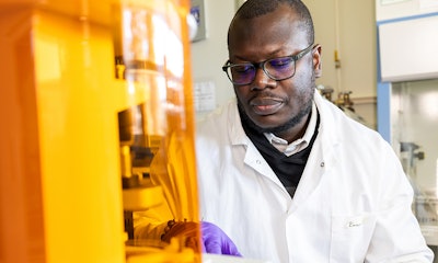
“To conduct our research and the testing that’s required — where we’re studying the mechanisms of complex lung diseases to eventually find new drug targets — we need to be able to make models that are comparable to human tissues,” said Dr. Osei, assistant professor in the Irving K. Barber Faculty of Science at UBC.
The scientists applied a bioink, composed of light-sensitive polymer-modified gelatin and a polymer called polyethylene glycol diacrylate, to 3D print a hydrogel that contains multiple cell types and channels to recreate vessels. The realistic model imitates the structure of a human airway, and the hydrogel functions much like the complex mechanical properties of lung tissue. The creation can improve how researchers study cellular responses to stimuli, said the university press release.
“Our goal was to create a more physiologically relevant in vitro model of the human airway. By integrating vascular components, we can better simulate the lung environment, which is crucial for studying diseases and testing therapeutics,” said Dr. Osei, who also works with UBC’s Centre for Heart Lung Innovation.
Dr. Osei and his colleagues performed multiple tests on the 3D model, including exposing it to cigarette smoke extract, which allowed them to observe markers of inflammatory responses to nicotine in lung tissue.

The study is a big step forward in assessing aspects of lung diseases, such as scarring and inflammation, and may lead to future cures for various illnesses. Being able to develop models that allow for testing is a significant advancement in respiratory research, Dr. Osei said.
“Our model is complex, but due to the reproducibility and optimal nature of bioprinting, it can be adapted to include additional cell types or patient-derived cells, making it a powerful tool for personalized medicine and disease modeling,” he said.