Myosteatosis measurements derived from artificial intelligence of coronary artery calcium (CAC) CT scans have shown potential in predicting future COPD risk.
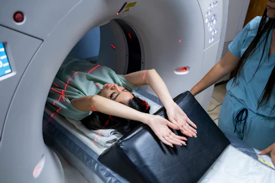
That’s according to the paper, “Artificial Intelligence-Derived Measurements From Coronary Artery Calcium CT Scan To Predict COPD: The Multi-Ethnic Study of Atherosclerosis,” published in Radiology: Cardiothoracic Imaging.

According to a press release, the study analyzed baseline CAC CT exams from 5,535 participants in the Multi-Ethnic Study of Atherosclerosis (MESA) and followed clinical outcomes for about 20 years. During follow-ups, 396 participants (7.1%) were diagnosed with COPD.
Investigators used a platform developed by HeartLung.AI called AI-CVD, which uses artificial intelligence to analyze CT screening and identify hidden risks, to identify myosteatosis — a CT marker of fatty infiltration and reduced muscle quality. They compared the predictive value of this measurement with another AI-derived emphysema-like lung measurement obtained from the same CT scans.

“The association between myosteatosis and COPD remained consistent across age, sex, obesity, smoking status and activity subgroups,” the authors wrote. This is the first study to investigate the association between AI-quantified myosteatosis and future COPD diagnosis.

“This is exactly where AI can change medicine,” said HeartLung.AI Founder and President Morteza Nachavi, MD. He explained that AI enables subtle quantitative findings, such as muscle fat infiltration, to be measured reproducibly from CT images even when they may not be practical to quantify visually during routine clinical interpretation.
The researchers stated that additional validation and studies are needed before myosteatosis can be established as a clinical COPD biomarker. Future studies are expected to examine additional populations, evaluate whether changes in muscle quality precede deterioration in pulmonary function and determine whether interventions that improve muscle quality may influence future COPD risk.