The National Heart, Lung and Blood Institute (NHLBI) awarded a five-year, $3 million R01 grant to University of Cincinnati researcher Nalinikanth Kotagiri, PhD. The grant will fund the study and development of an innovative imaging method that can provide real-time results. This momentous advancement will speed up the diagnosis and treatment of lung infections in critically ill patients.

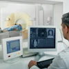
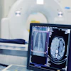
Dr. Kotagiri, an associate professor of pharmaceutical sciences at the UC James L. Winkle College of Pharmacy, will lead the federally funded study. He and his team will analyze the validity of injectable probes used during a PET (positron emission tomography) scan. The metallic contrast agents would amass at the infection site and immediately light up, signifying the infection’s presence, location and type.
“Our solution is to use imaging to identify what is causing the pneumatic episode within hours, to hasten a treatment plan,” said Dr. Kotagiri.
The current process to diagnose pneumonia and other lung infections contains several barriers. Chest X-rays alone cannot identify the nature of an infection—bacterial, viral or fungal. This requires a pathologist to capture and culture a lung tissue specimen by means of a bronchoscopy to determine specifics of the infection.
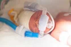
Scheduling and completing this invasive procedure followed by obtaining and reading its results can take a minimum of several days. This is lost time that patients do not have, said Kotagiri, especially those with underlying conditions like COPD or infectious pneumonia.
The study will use animal models to specifically assess bacterial and viral pneumonias in combination with COPD. However, the new imaging method would also theoretically apply to fungal pneumonia and other lung infections in conjunction with conditions such as cystic fibrosis.

Dr. Kotagiri also shared that the process to develop the contrast agent should be swift and straightforward. This would shorten the preparation time in the lab and allow for an earlier implementation in the clinical setting.
In addition to initiating a quicker treatment plan, Dr. Kotagiri said the new process could help clinicians improve treatment efficacy by prescribing an antibiotic that targets the pathogens identified in the imaging. Providers can also repeat the imaging following treatment to evaluate a patient’s response and ensure the infection is no longer present.