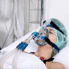
A recent study out of Germany and the Netherlands shares initial findings of a new CT protocol that concurrently evaluates lung structure and function. The limited study, published in Radiology on July 11, 2023, reports on early experience of using the same CT equipment paired with advanced software that amplifies the scale of the test using photon-counting CT (PCCT).

This comprehensive protocol was not previously possible with standard CT, which could primarily assess lung structure. The recently developed PCCT technology enables high image quality at a lower radiation dose and provides improved spatial resolution and spectral imaging options. Thus, the PPCT provides clinicians with an all-inclusive procedure that measures lung structure as well as lung function, particularly perfusion, ventilation and vasculature of the parenchyma.
The study indicates promising benefits over the standard chest CT procedure, which is the prevailing imaging tool to identify lung disease and monitor changes.
“The improvement in the contrast-to-noise ratio and spatial resolution of the pulmonary blood volume images was substantial,” said senior author Hoen-oh Shin, MD, professor of radiology at the Institute of Diagnostic and Interventional Radiology at Hannover Medical School in Hannover, Germany. “In my opinion, the most important advantage is the significantly improved spectral resolution, which enables new applications such as functional imaging of the lungs with CT.”
As part of the new protocol, researchers administered an intravenous contrast agent. They prompted patients to breathe in while completing an inspiratory PCCT, then five minutes later prompted patients to breathe out while completing an expiratory PCCT. Dr. Shin and colleagues attained an 85% success rate in attaining all CT-derived parameters.

“With the proposed protocol, we have also been able to answer many other questions related to post-COVID-19 condition, such as the detection of acute and chronic pulmonary emboli on CT angiography, and we are currently investigating whether perfusion changes can be quantified in microvascular damage or inflammatory areas,” Dr. Shin said.
The photon-counting CT protocol will provide clinicians with the complete data to recognize and analyze pulmonary impairment. It also has potential to improve lung imaging applications in surgery during the preoperative and postoperative stages. For example, clinicians could isolate areas of emphysema in patients who have chronic thromboembolic pulmonary hypertension. PCCT could assist in calculating surgical success and evaluating pulmonary pathology results in patients who have COPD or have had lung or stem cell transplant procedures.
The study, performed between Nov. 2021 and June 2022, included 196 patients with clinically indicated CT for various known and unknown lung function impairment. Additional studies will focus on refining mechanics, increasing processing time and enhancing the procedure’s capabilities. This innovation will create an updated, comprehensive modality for pulmonary evaluation that is proficient and efficient.