A deep learning model that uses a single inhalation lung CT scan has been shown to accurately diagnose and stage COPD, according to a study published in Radiology: Cardiothoracic Imaging.
COPD is typically diagnosed with a spirometry — or pulmonary function — test, which measures lung function through the quantity of air that can be inhaled and exhaled along with the speed of exhalation. CT images are also used to aid in COPD diagnoses. These often require two images — one taken at full inhalation and one at normal exhalation.

 Kyle A. Hasenstab, PhDSan Diego State University
“Although studies have recently shown that lung structure, quantitively measured using lung CT, can supplement COPD severity staging, diagnosis and prognosis, many of these studies require the acquisition of two images,” he said. “However, this type of protocol is not standard across institutions.”
Dr. Hasenstab, who is also assistant professor of statistics and data science at San Diego State University in California, said some hospitals are unable to implement expiratory CT protocols due to the added training required.
“[These] may not be feasible at many institutions due to the need for technologist training to acquire the images and radiologist training to interpret the images,” he said.
Another problem with these existing methods, said Dr. Hasenstab, is some elderly patients with impaired lung function struggle to hold their breath, which can impact the quality of CT images and the accuracy of the diagnosis.
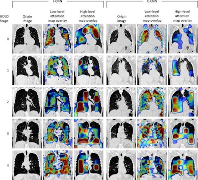
The researchers hypothesized that a single inhalation CT acquisition combined with a convolutional neural network (CNN) and clinical data would be sufficient for COPD diagnosis and staging. A CNN is a type of artificial neural network that uses deep learning to analyze and classify images.

The study gathered inhalation and exhalation lung CT images and spirometry data from 8,893 patients from 2007 to 2011. The average age was 59 years and all of them had a history of smoking. Researchers used the data to train the CNN to predict spirometry measurements using clinical data and either a single- or multi-phase lung CT.
The spirometry predictions were then used to predict the Global Initiative for Obstructive Lung Disease (GOLD) stage of the disease. The results of the study showed that a CNN model developed using only a single respiratory phase CT image accurately diagnosed COPD and was also accurate within one GOLD stage. When clinical data were added, the CNN model’s predictions were even more accurate.
“Although many imaging protocols for COPD diagnosis and staging require two CT acquisitions, our study shows that COPD diagnosis and staging is feasible with a single CT acquisition and relevant clinical data,” said Dr. Hasenstab. “Reduction to a single inspiratory CT acquisition can increase the accessibility to this diagnostic approach while reducing patient cost, discomfort and exposure to ionizing radiation.”