Researchers from Vanderbilt University and Vanderbilt University Medical Center (VUMC) have developed three-dimensional video images of lab-grown mouse lung tissue to better understand pulmonary resilience. Their findings, “Epithelial Outgrowth Through Mesenchymal Rings Drives Lung Alveologenesis,” were published in the American Society for Clinical Investigation journal, JCI Insight.

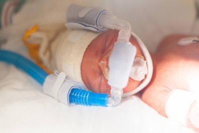
The groundbreaking knowledge allows scientists to get a closer look at how damaged or underdeveloped lung tissue heals and regenerates, with the goal to improve prevention and treatment of bronchopulmonary dysplasia (BPD) — a lung condition that occurs in approximately half of premature babies who are born two to four months early.
“For the first time, we’ve been able to live-image the lung as it forms and quantify and measure those cellular movements that come together to make an organ with a surface area large enough for gas exchange,” said senior author Jennifer Sucre, MD, in a university news release. Dr. Sucre is associate professor of pediatrics and cell and development biology, as well as director of the Biodevelopmental Origins of Lung Disease (BOLD) Center at VUMC.
Infants who have BPD require oxygen and mechanical ventilation to help them breath. However, this therapy can also further damage their immature lung tissue. Even after ventilation ends, the babies are at risk of developing long-term respiratory problems, such as COPD.
“If we can understand how the lung forms, then we have a blueprint for how to grow new lungs after injury,” said first author Nick Negretti, PhD, senior postdoctoral fellow in the Sucre lab.
The group of researchers used four-dimensional microscopy, a technique called scanned oblique plane illumination (SOPi), to analyze images of living neonatal mouse lungs. This led to the discovery that alveolar epithelial cells grow from mesenchymal rings made up of myofibroblasts and other cells that stimulate tissue formation.

The innovative, 4D technical approach supports testing and identification of specific molecules and pathways that control alveoli development.
“In more than 2,000 hours of live imaging, we never saw a single instance of ingrowing septation. The structures that we previously thought were ingrowing septal tips were artifacts of traditional two-dimensional imaging,” Dr. Negretti said.
 Jennifer Sucre, MDVanderbilt University
“Getting the model right and understanding how the cells move changes the questions we’re asking,” Dr. Sucre said. “Instead of looking at cells that divide alveoli, we’re now looking for the pathways that guide the cells’ outward growth and help them find each other.”
The model also provides a solution to assess lung development at the cellular level and understand how genetics, environmental exposures and pharmacological therapies, particularly those used in a neonatal intensive care unit (NICU), impact it.

Dr. Sucre said she and her lab team are “keen to understand the cellular behaviors and dynamics in the more resilient mouse. What are the pathways in the resilient lung that can repair it after infection and injury? How do we bottle that?” This knowledge, she added, could help develop future treatments that don’t interfere with lung maturation.
“Mice have an extraordinary ability to repair the lung. I want to give babies the superpower of the mouse,” she said.